
Ocular Imaging
Retinal imaging
As part of your diagnosis, your ophthalmologist will need to photograph your retinas.
As well as confirming the diagnosis, the images will prove useful in planning your treatment. There are several different ways of taking pictures of the retina.
Fundus photography
A fundus camera is a special camera used to take photographs of the inside of your eye. It can capture high resolution colour images of your macula.
Fluorescein angiography
Angiography ('the dye test') is an examination that creates detailed images of your blood vessels and the bloodflow inside them. A special dye is injected into your blood vessels and pictures are taken that show any abnormalities inside them.
The procedure is useful for various retinal conditions including macular degeneration, diabetic retinopathy and retinal vein thrombosis.
Your ophthalmologist will inject a special dye called fluorescein into a vein in your arm. The dye will move through your blood vessels into your retina. They will take a series of pictures using a special camera.
These images will allow your ophthalmologist to confirm diagnosis and plan treatment.
Indocyanine green (ICG) angiography
The technique used for indocyanine green (ICG) angiography is the same as for fluorescein angiography, but the dye is different. ICG dye can highlight slightly different problems in your eyes.
Optical coherence tomography (OCT)
Optical coherence tomography (OCT) uses special rays of light to scan your retina and produce an image of it. This can provide detailed information about your macula. For example, it will tell your ophthalmologist whether your macula is thickened or abnormal, and whether any fluid has leaked into the retina.
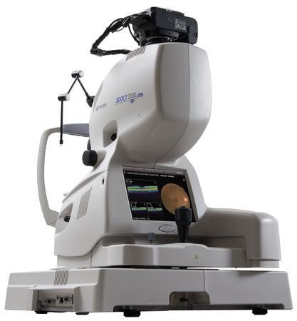 Topcon 3D OCT-2000
There are two high quality spectral domain OCT scanners available at our facility:
The Topcon 3D OCT-2000 incorporates a high resolution fundus camera and
the Spectralis OCT integrated with scanning laser ophthalmoscopy and blue laser autofluorescence.
Both scanners provide detailed images of the structure of the various layers of the macula and the spectralis additionally gives information about function through autofluorescence (AF)
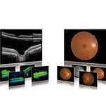 Images with Topcon 3D OCT scanner
 Spectralis image of macula